Chapter 3
Advancing cancer research and improving patient care are currently hindered by several major challenges. In general, these challenges are rooted in our incomplete understanding of tumor heterogeneity and emergence of treatment resistance. These gaps in knowledge limit our ability to prognosticate tumor progression and stratify patients, resulting in high clinical trial failure rates and delayed access to new life saving treatments, and ultimately significant financial burden and poor clinical outcomes for patients (Figure 2, top panels).
Although the challenges encountered in oncology tend to follow a linear cause-and-effect process, multiomics is not constrained by this framework. Rather, multiomics has emerged as a tool capable of addressing the challenges found at every level. For example, integrated omics datasets can reveal deep biological insight into tumor heterogeneity and mechanisms of treatment resistance. Harmonized datasets can identify novel biomarker signatures to not only determine which patients would be most likely to respond to treatment but also provide insight into the mechanisms that regulate treatment response. Multiomics can also significantly increase the likelihood of success for clinical trials due to the level of detail that it reveals about therapeutic target specificity, mechanisms of action, toxicity, and patient stratification, all of which lead to more efficient drug development, better patient outcomes, and lowered healthcare costs (Figure 2, bottom panels).
In this chapter, we will evaluate three case studies that demonstrate the capabilities of multiomics in helping to address key challenges in cancer research. We hope these studies inspire the use of integrative multiomics as a staple in future cancer studies.
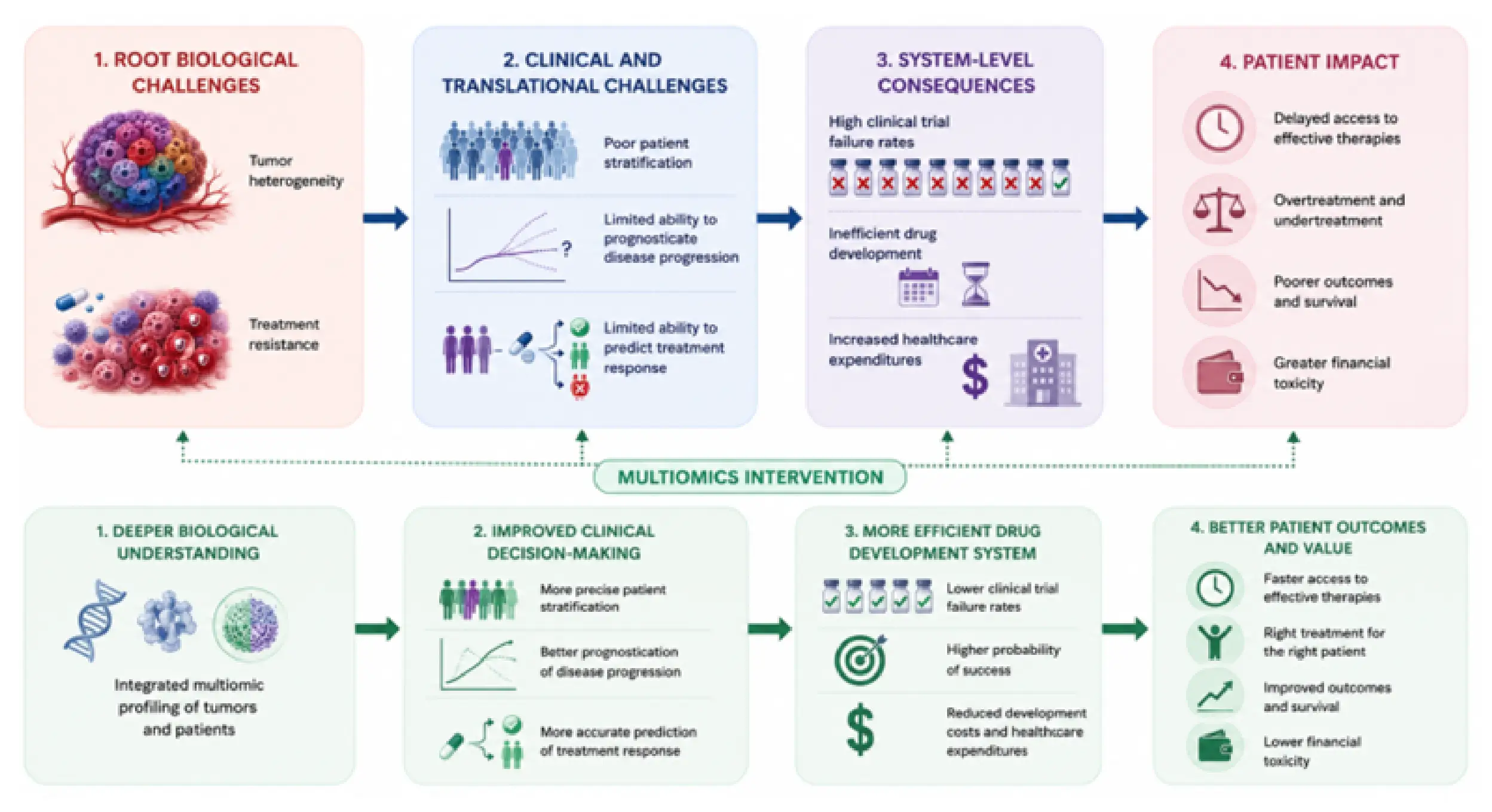
Figure 2. A summary of challenges that hinder advancement of cancer research and patient care, and how multiomics may address the challenges found at all levels. Image licensed under CC BY 4.0.
Cancer patients suffer greatly from a lack of biomarkers to help predict clinical outcomes and select the most effective treatment strategies before therapy begins. Patients with hepatocellular carcinoma (HCC) often experience highly variable disease progression, making accurate prognosis particularly beneficial to this patient population. Previous studies had shown that transcriptomic signatures could identify molecular subtypes of HCC associated with good or poor survival, but these approaches required invasive biopsy procedures. In this study, investigators hypothesized that this same prognostic information could be captured in a blood sample. Since expression of tumor oncogenes disrupts metabolism to drive tumor progression, investigators approached the study objective by analyzing integrated metabolomic and transcriptomic datasets.
Metabolon’s HD4 platform was used to perform global metabolomics analyses on paired tumor and adjacent non-tumor tissues on an Asian HCC discovery cohort and an independent validation cohort of 408 patients. Metabolic genes were identified from the Human Metabolic Reaction database. Untargeted metabolomics was performed using Metabolon’s Discovery HD4 platform, and principal component analysis was used to distinguish tumor from adjacent non-tumor tissues based on global metabolite profiles. Differential metabolic genes and metabolites were identified using paired t-tests with Benjamini-Hochberg false discovery rate correction, after which a novel two-step Spearman correlation analysis combined with 10,000 permutation tests linked metabolic gene expression to tumor metabolites and subsequently tumor metabolites to serum metabolites, identifying 491 metabolic genes, 40 tumor-specific metabolites, and 75 prognostic serum metabolites.
Consensus clustering independently classified patients using each molecular dataset, while subclass mapping validated these classifications against independent cohorts and previously established HCC molecular subtypes (Figure 3). Kaplan-Meier survival analysis, log-rank testing, and Cox proportional hazards modeling demonstrated that transcriptomic, tissue metabolomic, and serum metabolomic data each identified the same poor prognosis patient subgroup (Figure 4), with serum metabolite classification producing a hazard ratio of 4.4 for overall survival and outperforming the conventional biomarker α-fetoprotein, which did not significantly discriminate outcomes. Finally, pathway analyses further revealed that the poor-prognosis subtype was characterized primarily by dysregulated lipid metabolism and fatty acid β-oxidation, with acylcarnitines comprising the dominant prognostic metabolite class.
Through the use of multiomics, circulating serum metabolites were shown to faithfully recapitulate the underlying molecular state of HCC and accurately classify patients into clinically meaningful prognostic subgroups. This approach has the potential to improve patient stratification, guide treatment selection, identify individuals who may benefit from more aggressive therapies or closer monitoring, and facilitate enrollment into precision medicine clinical trials using a minimally invasive blood test instead of surgically obtained tissue. Following further clinical validation, serum metabolite panels could provide a more accessible and clinically practical means of prognosis than current molecular profiling methods.
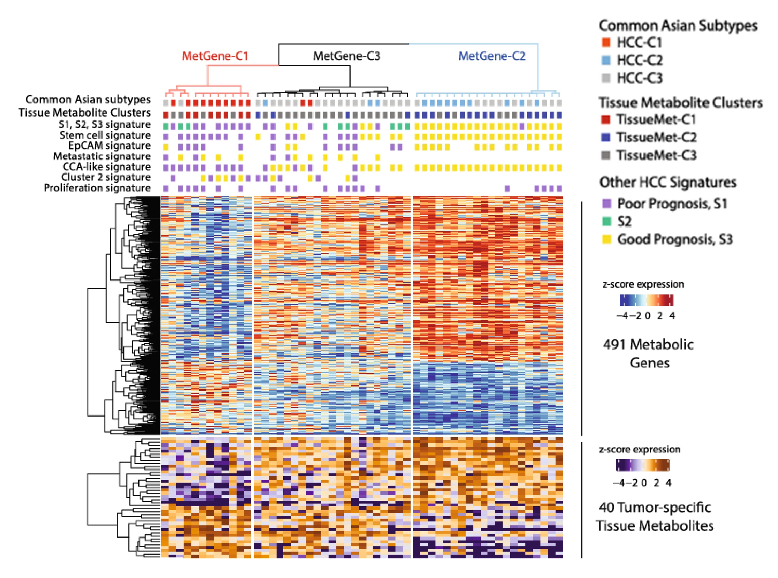
Figure 3. Correlation analysis of tumor metabolic genes and tissue metabolites. Heatmap of gene expression from 491 metabolic genes in the tumor tissues from HcC patients. The top panel shows clusters of patients based on 1) consensus clustering from 491 metabolic genes identified through permutation test, 2) previously identified Asian subtypes, and 3) tissue metabolite (TissueMet) clusters, which are based on consensus clustering from 40 tumor-specific metabolites. The bottom panel shows two heatmaps based on z-score expression of metabolic genes and tumor-specific tissue metabolites. Image licensed under CC BY 4.0.
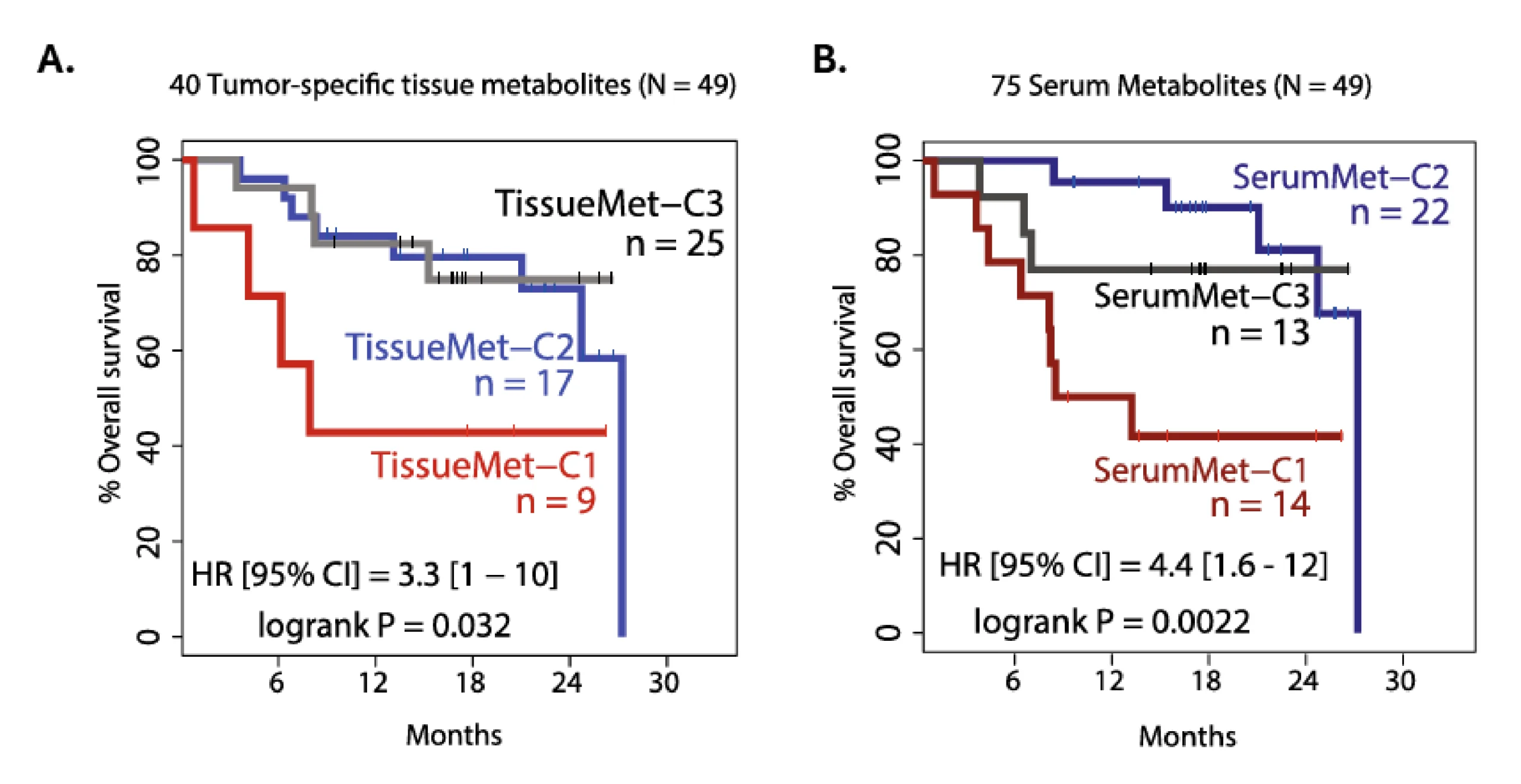
Figure 4. Kaplan-Meier plots showing survival probabilities of HCC patients according to consensus clustering based on abundance of (A) 40 tumor-specific metabolites, and (B) 75 serum metabolites. Image licensed under CC BY 4.0.
Treatment resistance represents a multi-level challenge in clinical oncology. Not only do resistant tumors complicate treatment decisions and often lead to poor patient outcomes, but they also require additional imaging, biopsies, molecular testing, and hospital stays, which significantly increases the cost of healthcare. Despite decades of research, there are still significant gaps in knowledge on the mechanisms that induce genetic adaptations and subsequent metabolic reprogramming that make tumors less susceptible to treatment. In the context of highly complex tumor biology, multiomics approaches are vital to both understanding the processes that drive treatment resistance and identifying biomarkers that could predict treatment response. Multiomics approaches have been particularly important to patients with clear cell renal cell carcinoma (ccRCC), where treatment resistance remains a major barrier to long-term disease control. In this study, investigators used multiomics approaches to characterize metabolic alterations associated with treatment resistance and subsequent progression of ccRCC31.
Global metabolomics profiling was performed on 40 primary ccRCC tumor samples and 20 noncancerous kidney tissues, while transcriptomic profiling was conducted using microarray data from 10 ccRCC tumors and matched normal kidney tissues (GSE47032 dataset). Analyses included principal component analysis (PCA) to compare metabolic profiles, metabolite and gene set enrichment analyses to identify dysregulated pathways, and fold-change analyses to rank altered genes. Kaplan-Meier survival curves and Cox regression models were used to evaluate associations between molecular markers and clinical outcomes and to determine whether NDUFA4L2 was an independent prognostic factor.
Untargeted metabolomic profiling identified 344 significantly altered metabolites and clearly distinguished ccRCC tumors from normal kidney tissue. The metabolic signature indicated that ccRCC cells reprogram glucose metabolism by diverting glucose away from mitochondrial oxidation and toward the pentose phosphate pathway (PPP), which generates NADPH and nucleotide precursors that support tumor growth and protect against oxidative stress (Figure 5). Tumors also exhibited evidence of impaired mitochondrial oxidative phosphorylation, including alterations in TCA cycle metabolites, increased glutamine and glutamate utilization, reductive carboxylation, and reduced ATP production, suggesting a shift toward alternative metabolic pathways.
Integrated transcriptomic analysis identified NDUFA4L2 as the most highly overexpressed gene in primary renal cancer cells. NDUFA4L2, a hypoxia-inducible factor (HIF-1) target gene, was linked to reduced oxidative phosphorylation, mitochondrial dysfunction, and adaptation to hypoxic tumor environments. Functional studies showed that NDUFA4L2 promotes tumor cell survival, migration, and resistance to chemotherapy, while gene silencing restored mitochondrial activity, increased oxidative stress, reduced cell viability, and enhanced sensitivity to cisplatin-induced apoptosis (Figure 6). Analysis of 390 patients further demonstrated that elevated NDUFA4L2 expression was associated with shorter cancer-specific and progression-free survival and remained an independent prognostic factor after adjustment for established clinical variables.
This study demonstrates how metabolomics can uncover clinically relevant metabolic subtypes of ccRCC and identify key drivers of tumor progression. The integrated analysis revealed a distinct metabolic phenotype characterized by enhanced glycolysis, increased pentose phosphate pathway activity, impaired mitochondrial oxidative phosphorylation, and over-expression of NDUFA4L2. These findings may improve patient stratification by enabling more accurate prognostication, earlier identification of aggressive disease, and selection of patients most likely to benefit from metabolism-targeted therapies. In addition, because NDUFA4L2 was independently associated with poorer survival outcomes and contributed to chemotherapy resistance, it represents a promising prognostic biomarker and therapeutic target that could support biomarker-guided clinical trials and reduce trial-and-error treatment approaches.
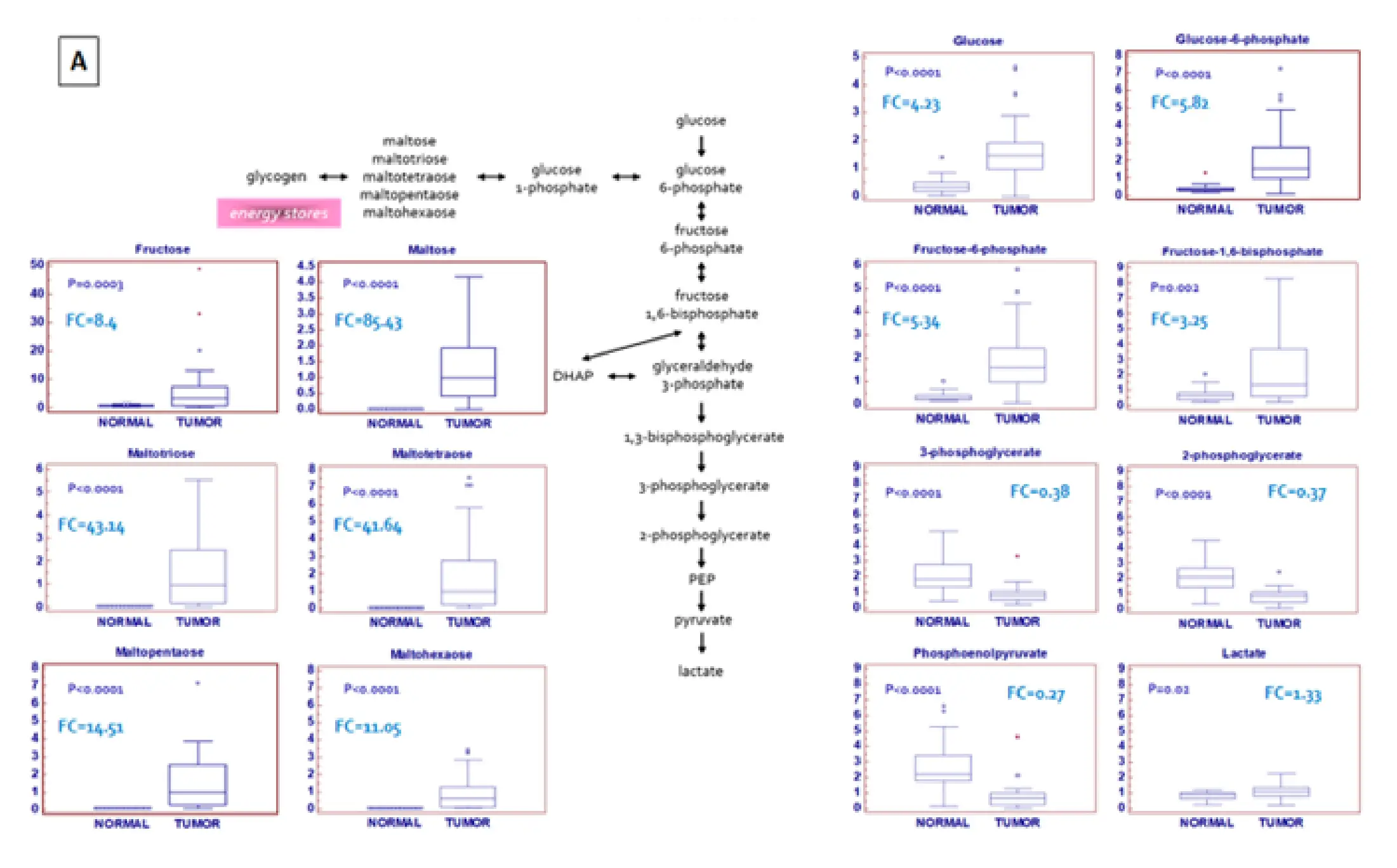
Figure 5. A schematic model summarizing the differences in glucose metabolism between normal and tumor tissue. Image licensed under CC BY 4.0.
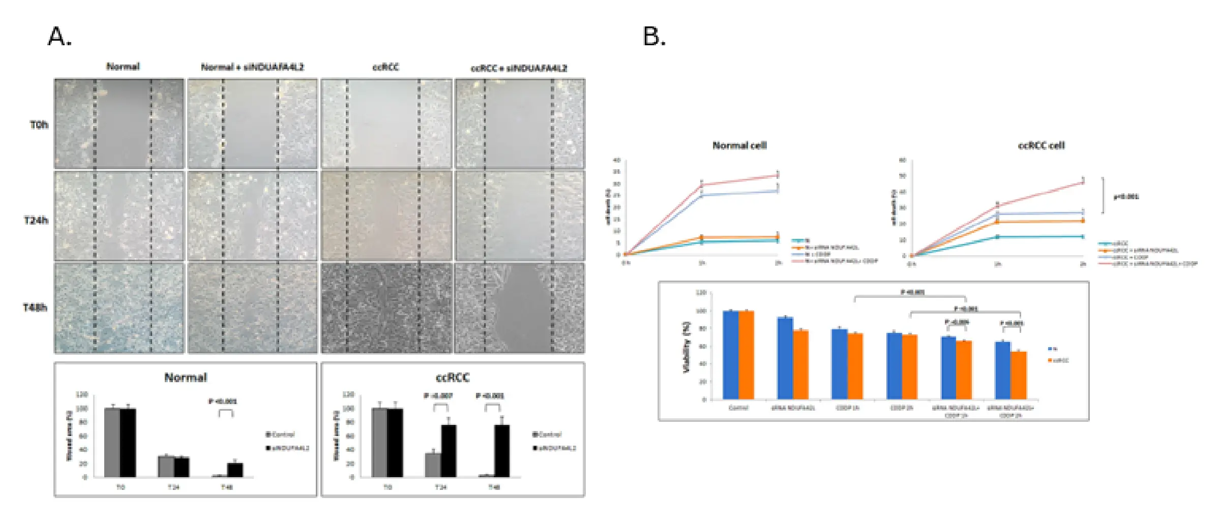
Figure 6. (A) Scratch wound assay showing that cell migration is decreased in response to the silencing of NDUFA4L2 expression. (B) NDUFA4L2 plays a role in resistance to cisplatin-induced cytotoxicity as shown by the significantly higher death rate of treated tumor cells with silenced NDUFA4L2 expression compared to cells with normal NDUFA4L2 expression. Image licensed under CC BY 4.0.
Despite advances in immuno- and targeted therapies, the ability to predict which patients will respond to treatment remains limited. In clinical practice, the absence of reliable predictive biomarkers often forces treatment decisions to rely on population-level evidence rather than individual tumor biology, potentially delaying the start of effective therapies and increasing healthcare costs. Unpredictable response to treatment may also dilute therapeutic effects in clinical trials by grouping responders and non-responders together, potentially causing effective therapies to fail to meet their primary endpoints.
As demonstrated in the following study, variability in response to immune checkpoint inhibitors (ICT) remains a major obstacle to optimizing outcomes in patients with metastatic melanoma32. Multiomics approaches can help address this challenge by integrating information from multiple biological layers to identify biomarkers that predict treatment response and guide patient stratification. The goal of this study was to determine whether the response to ICT therapy could be predicted based on the composition of the gut microbiome and gut metabolome prior to the start of treatment.
The study used integrated metagenomic and metabolomic analyses of pretreatment stool samples from 39 patients with metastatic melanoma receiving ICT. Shotgun metagenomic sequencing was performed to characterize gut microbial composition at the species level and assess microbial functional pathways, and untargeted LC-MS metabolomics was used to identify metabolites associated with treatment response and evaluate links between microbial metabolism and therapeutic efficacy. Statistical analyses included linear discriminant analysis effect size (LEfSe) to identify microbial taxa associated with response status, hierarchical clustering to assess microbiome stability over time, LEfSe and Kruskal-Wallis testing to compare microbial functional pathways, and Welch’s two-sample t-tests to identify metabolites that differed between responders and non-responders.
Metagenomic analysis identified several bacterial species that were enriched in patients who responded to ICT therapy. Across all treated patients, responders showed higher levels of Bacteroides caccae and Streptococcus parasanguinis. Additional response-associated species varied by treatment regimen, with Faecalibacterium prausnitzii, Bacteroides thetaiotaomicron, and Holdemania filiformis enriched among patients receiving ipilimumab plus nivolumab, and Dorea formicigenerans enriched among pembrolizumab responders. Despite these differences in microbial composition, overall gut microbial diversity did not differ significantly between responders and nonresponders, suggesting that specific microbial species may be more important than overall diversity in influencing treatment outcomes. Functional metagenomic analyses further showed that responder microbiomes were enriched for pathways involved in fatty acid biosynthesis and inositol phosphate metabolism, indicating that microbial metabolic activity may contribute to immunotherapy efficacy.
Untargeted metabolomics revealed substantial differences in fecal metabolite profiles, with 83 metabolites significantly associated with treatment response. One of the strongest associations involved 15:2 anacardic acid, a plant-derived metabolite found in foods such as cashews and mangos. Levels of this metabolite were approximately 62-fold higher in responders across all ICT-treated patients and approximately 94-fold higher in responders receiving ipilimumab plus nivolumab (Figure 7). Most individuals with the highest levels reported regular cashew consumption, suggesting a potential link among diet, the gut metabolome, and response to immunotherapy.
This study demonstrates how metabolomics can identify biochemical signatures associated with immunotherapy response, highlighting 15:2 anacardic acid as a strong predictor of treatment efficacy. Because anacardic acid is known to enhance innate immune responses, its association with favorable outcomes suggests a potential biological mechanism through which the gut metabolome may influence ICT effectiveness and raises the possibility of dietary interventions to improve treatment response.
More broadly, the findings illustrate how multiomics can improve patient stratification by distinguishing likely responders from nonresponders before treatment begins, potentially reducing exposure to ineffective therapies and lowering healthcare costs. The study also expands the precision oncology paradigm beyond tumor biology, demonstrating that host factors such as the gut microbiome and metabolome may play a critical role in determining therapeutic outcomes and could help explain why patients with similar tumors often experience markedly different responses to treatment.
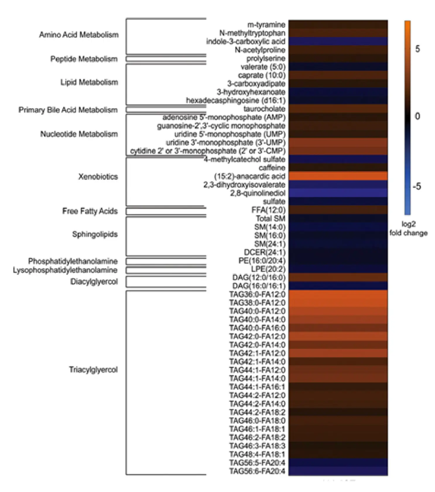
Figure 7. Unbiased metabolomics analysis of stool metabolites from melanoma patients prior to treatment with ICT (n = 39). The heat map shows the normalized relative abundance of metabolites comparing responders to those with progressive disease. Orange = fold increase; blue = fold decrease. This analysis identified (15:2)-anacardic acid as a strong predictor of treatment efficiency. Image licensed under CC BY 4.0.
Once you see the full value of metabolomics, the only remaining question is: who does it best? While many laboratories have metabolite profiling or analytical chemistry capabilities, comprehensive metabolomics technologies are extremely rare.
Accurate, unbiased metabolite identification across the entire metabolome introduces signal-to-noise challenges that very few labs are equipped to handle. Also, translating massive quantities of data into actionable information is slow, if not impossible, for most because proper interpretation takes two things that are in short supply: experience and a comprehensive database.
Using our robust platform and visualization tools, our experts are uniquely able to tell you more about your molecule and develop assay panels to help you zero in on the results you need.
Coverage
Ability to interrogate thousands of metabolites across diverse biochemical space, revealing new insights and opportunities
Comparability
Ability to integrate the data from different studies into the same dataset, in different geographies, among different patients over time
Competency
Ability to inform on proper study design, generate high‐quality data, derive biological insights, and make actionable recommendations
Capacity
Ability to process hundreds of thousands of samples quickly and cost‐efficiently to service rapidly growing demand
Request a quote for our services, get more information on sample types and handling procedures, request a letter of support, or submit a question about how metabolomics can advance your research.
Corporate Headquarters
617 Davis Drive, Suite 100
Morrisville, NC 27560
Mailing Address
P.O. Box 110407
Research Triangle Park, NC 27709
Phone
+1 (919) 572-1711
Fax
+1 (919) 572-1721
International Headquarters
Metabolon GmbH
Zeppelinstraße 3
85399 Hallbergmoos
Germany